Contact

+55 (11) 9 9686 9874
Yuri de Stefani


Master's Degree Project
Computed Tomography (CT) is a technique that allows the acquisition of three-dimensional images of the human body. Since its introduction in clinical practice, this imaging modality has been essential for disease diagnosis and is used worldwide. In recent decades, dosimetric evaluation in CT has been performed using the physical quantity. However, with the evolution of CT equipment and the use of wider beams, the use of the physical quantity has become inadequate as it underestimates the dose distributed along the longitudinal axis of simulated objects. Thus, a new measurement methodology and dose representative physical quantities for CT equipment were proposed. This methodology involves determining dose distributions for different scan lengths, , represented by equilibrium approximation functions, h(L). With these functions, the physical quantities equilibrium doses (Deq), equilibrium lengths (Leq), and scatter fractions (alfa) are estimated. The methodology also allows for the estimation of geometric efficiency in the z-direction. In this study, this methodology was implemented using CT equipment at the Radiology Institute of Hospital das Clínicas FMUSP (GE Discovery CT750HD from General Electric, Aquilion CXL 128 from Toshiba, and Brilliance 64 from Philips). Measurements were taken using two data acquisition methods (single scan and serial). The measurements were performed using different protocols for each equipment, varying the pitch, voltage, filter, and rotation axis. The curves obtained by both methods were compatible. The single scan method was chosen to estimate parameters and the curve because it has lower operational costs and provides a more complete curve than the serial method.
(That was the abstract of my dissertation)
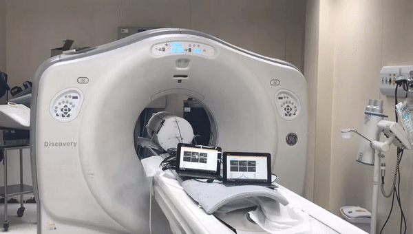
The following image shows the phantom build.

The following giff shows the phantom been measured.